Glaucoma – What is glaucoma?
Green cataract is a disease of the optic nerve that, if left untreated, can lead to vision loss. Here you will find compact: early signs, meaningful examinations and effective therapies.

What is glaucoma? – Definition & Shapes
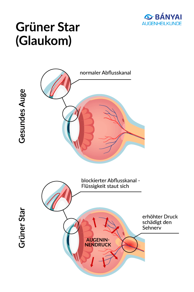
Green cataract, medical glaucoma, refers to several eye diseases that damage the optic nerve – initially often without pain. The balance of aqueous humor production (in the ciliary body) and aqueous humor drainage via the angle of the chamber with trabecular mechanism is decisive.
If it is out of balance or if there are circulatory disorders on the optic nerve/retina, the risk of vision loss increases. Early clarification helps to slow down the course. The name “Green Star” describes the blue-green shimmer of the iris, on the other hand also the “fixed gaze” in the blindness of the eye.
Since glaucoma usually begins gradually, it helps to recognize the first signs of the cataract. For example, minor failures in the peripheral area of the visual field can be an indication at first. They usually appear as a blind spot that affects vision.
Glaucoma is one of the most common irreversible causes of blindness in Europe. Green Star surgery can provide a remedy.
30 years of experience and more than 55,000 treatments – Welcome to Bányai Augenheilkunde!
Whether you have vision problems, an eye condition, or are simply seeking preventive care, my team and I are dedicated to addressing your needs at our multiple locations in the Stuttgart and Karlsruhe areas. When you choose to receive treatment from me, you’ll benefit from comprehensive diagnostics and consultation, state-of-the-art laser procedures, and 30 years of experience.
What forms of glaucoma exist?
The most common form of glaucoma is primary open-angle glaucoma – which occurs gradually and with little discomfort. Beneficial factors for this form of glaucoma include severe farsightedness or myopia.
Secondary glaucoma
Secondary glaucoma (secondary open-angle glaucoma) arises as a result of a pre-existing condition – such as diabetes mellitus, farsightedness, inflammation or after eye injuries – and sometimes as a side effect of steroids; treatment depends on the cause.
Narrow angle glaucoma
In narrow-angle or angle-block glaucoma, the ventricular angle is too narrow or blocked, the drainage is acutely blocked: there is a risk of a glaucoma attack with visual disturbances, severe pain and nausea – here the eye clinic is immediately the right place.
Normal pressure glaucoma
In normal pressure glaucoma, optic nerve damage occurs despite “normal” intraocular pressure, often due to vascular and circulatory disorders. Rare, congenital forms – such as primary congenital glaucoma – occur in childhood.
Green cataract symptoms – detect early signs?
Green cataract symptoms are often unremarkable at first – glaucoma usually does not cause any pain at first. Pay attention to light sensitivity, halos (coloured circles around light sources), uncertain vision at dusk and small visual field gaps at the edge.
These indications do not prove glaucoma, but are a clear signal for early detection and a thorough diagnosis (measuring intraocular pressure, OCT, visual field test) by the ophthalmologist.
- Visual field defects or “blind spots” (especially on the side)
- Eye pain (rare in early stages), headache, nausea
- red eyes, eye tears, dilated pupils
- Decrease in visual acuity, poorer color contrasts
- Light sensitivity and halos around lamps
Emergency – acute glaucoma attack
Sudden severe headache/eye pain, red eye, vision loss, very hard eyeball or nausea speak for an acute glaucoma attack. This is a medical emergency with a risk of blindness – please act immediately (medical assistance/emergency service).
Important note: Individual complaints (e.g. headaches only) do not automatically mean glaucoma. If several symptoms coincide or there are doubts, have the intraocular pressure, OCT and visual field checked by your ophthalmologist.
What should your next steps be? Make an appointment for glaucoma clarification and carry out a diagnosis of cataracts (OCT & visual field) at the ophthalmologist. In the event of a glaucoma attack, book an appointment with the ophthalmologist immediately!
If you are interested in Green Star surgery, you will find all the information on ophthalmologist Bányai.
Green Star Causes & Risk Factors
Green cataract causes usually depend on the interaction of aqueous humour production in the ciliary body and the aqueous humour outflow via the angle of the chamber/trabecular structure. The ventricular angle is formed inside the eye by the cornea and iris; if the outflow is disturbed or if there are circulatory disorders on the optic nerve/retina, the risk of vision loss increases.
The intraocular pressure is measured in mmHg and is often between 10–21 mmHg – but your individual target value is decisive. The measurement (tonometry) is central to this and is always evaluated in context (including the influence of corneal thickness, time of day).
The risk of glaucoma increases from 60+, with family history, severe myopia, diabetes, sleep apnea, blood pressure fluctuations and with long-term cortisone therapy. In normal pressure glaucoma, vascular factors and nocturnal drops in blood pressure play a special role.
Glaucoma early detection and prevention – what’s the point?
Regular checks enable early detection of glaucoma and are the best protection: measuring intraocular pressure (tonometry, mmHg), OCT (RNFL) and visual field show changes early.
Adjust systemic factors well (e.g., blood pressure, diabetes, sleep apnea), discuss cortisone therapies, and apply glaucoma drops correctly. In this way, we achieve the individual target value and keep the optic nerve stable in the long term.
Glaucoma diagnosis & examinations: intraocular pressure, OCT & visual field
Glaucoma diagnosis is based on the combination of measuring intraocular pressure (tonometry, mmHg), OCT of the optic nerve/retina (RNFL) and visual field test (perimetry).
The decisive factor is not a single value, but the overall picture – including corneal thickness, time of day and course. From all findings, we set your target value and plan the control intervals.
Measuring intraocular pressure (tonometry)
The measurement of intraocular pressure (tonometry, mmHg) is central – reference values are often between 10–21 mmHg. “Too high” is what is above your target value. Methods: Goldmann‑Applanation (Gold‑Standard), Non‑Contact (Air Blast) or iCare (Rebound).
In the case of fluctuating values, a daily pressure profile makes sense; the corneal thickness (pachymetry) influences measurements.
Oct (RNFL) – Detect optic nerve early
The OCT shows the thickness of the nerve fiber layer in micrometers and makes the loss of nerve cells visible early.
This is how we detect early damage and assess the progression over time (standard-data-comparison, trend analysis). The examination is contactless and takes only a few minutes.
Visual field test (perimetry)
Perimetry shows functional failures (e.g. arcuate defects). Programs such as 24-2 or 30-2 are common. Since there is a learning curve, results in question are repeated in a timely manner. For driver’s license and professional requirements, the result is often relevant.
Brief conclusion: A reliable diagnosis is only made from pressure, OCT and visual field. This is how we define realistic target values and the appropriate therapy.
ONLINE Glaucoma Surgery Eligibility Test
Are you wondering if you’re a good candidate for glaucoma surgery? Take our online aptitude test now and find out your results in just a few clicks.
Glaucoma treatment & therapy – individual after diagnosis
The glaucoma treatment is based on lowering the intraocular pressure. After the glaucoma diagnosis, we jointly choose the therapy that reliably reaches your targetintraocular pressure and fits into everyday life.
At the Bányai Eye Centre, we rely on a clearly structured step strategy – with a focus on effective pressure reduction and reliable aftercare (controls with OCT and visual field).
Eye drops for glaucoma – start if useful
Eye drops can reduce aqueous humor production in glaucoma or improve drainage. Preparations (e.g. prostaglandin analogues such as latanoprost) are selected individually and checked regularly.
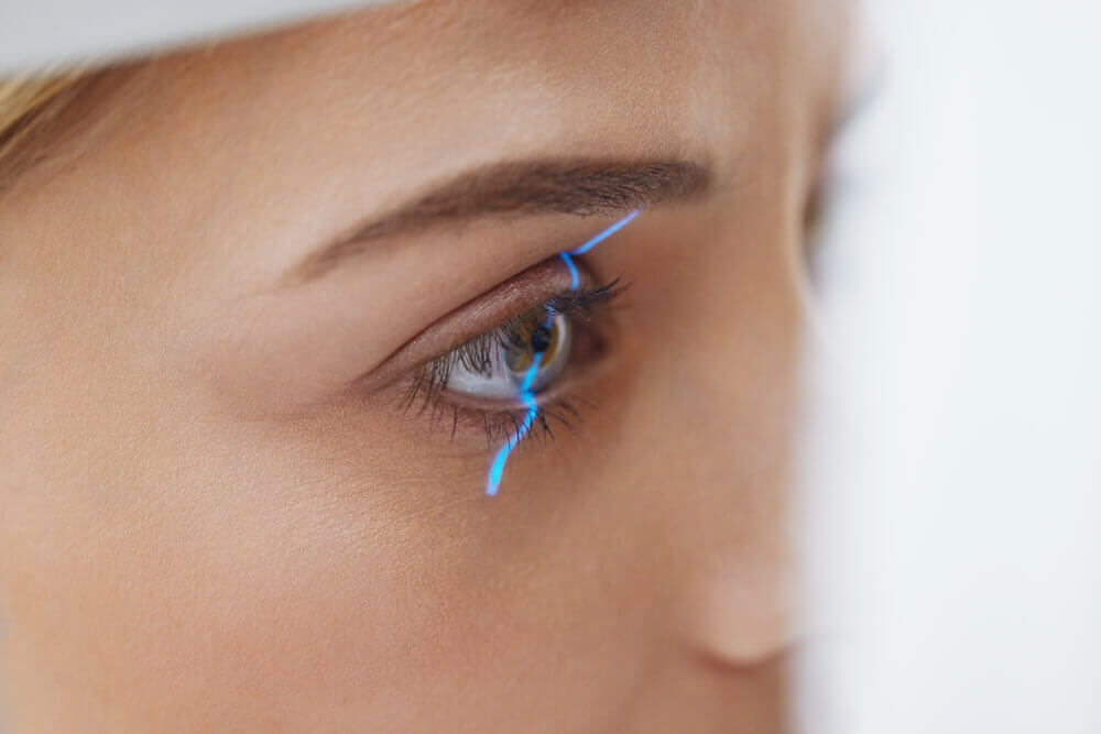
SLT laser – gentle improvement
Selective laser trabeculoplasty (SLT) improves the outflow in the trabecular structure and thus lowers the pressure. The procedure is performed on an outpatient basis, is well tolerated and can be repeated if necessary. In many patients, the effect is comparable to first-line therapy with drops – without daily use.
Glaucoma surgery – effective pressure reduction
If drops/SLT do not safely reach the target value, we recommend a glaucoma surgery. In the Bányai eye centre, trabeculectomy and – if there is also relevant lens opacity – cataract surgery are used.
Trabeculectomy is an established procedure with strong pressure reduction and close follow-up. Cataract surgery can measurably reduce intraocular pressure and is suitable if cataracts impair visual quality and diagnostics anyway; this can improve visual performance and often support pressure targets at the same time. We currently do not use the MIGs procedure.
What does this mean for you?
- The goal is a sustainable pressure reduction with the most effective and appropriate method.
- We offer SLT lasers on site; MIGS do not.
- In case of advanced glaucoma or insufficient effect, trabeculectomy is the most compressive option.
- If there is also a cataract, its operation can favour an IOP reduction – we will discuss whether this makes sense in your case.
Laser Eye Surgery at the Bányai Laser Eye Center
Book your personal consultation online now—with no obligation!
Laser Eye Surgery, Diagnostics & Consultations on Ophthalmology at the Bányai Laser Eye Center – Dr. Liliana Bányai

Julia M from Böblingen
Review from 07.08.2025
I received an appointment by email very quickly and was able to choose a suitable time flexibly – that was super uncomplicated. The entire staff…
more
David V from Ditzingen
Review from 15.07.2025
Staff is very helpful. Got an appointment very quickly (by email). I was able to choose an appointment very flexibly. All in all, great experience.
more
Carsten G from Böblingen
Review from 10.06.2025
I recently had my eyes lasered successfully here. Everything just worked wonderfully, I can finally see without glasses without any problems, for me it was…
more
Edith L from Weil der Stadt
Review from 27.12.2024
Fortunately, I was able to undergo a laser treatment, because the result is impressive. The procedure was short and painless and the treatment very pleasant.
more
Ulrich H from Böblingen
Review from 11.08.2024
Great address when it comes to eye lasers. I am completely satisfied and highly recommend it.
more
Benjamin H from Stuttgart
Review from 11.06.2024
That’s how a doctor works. Pretty easy to get an appointment, everything supported digitally… and then everyone was friendly and helpful.
more
Marion G from Botnang
Review from 31.01.2024
I am totally enthusiastic about my new eye and am looking forward to the second surgery. Yesterday I drove in the dark to refuel, because…
more
Claudia from Leonberg
Review from 22.10.2023
Great doctor, great team, the Relex Smile surgery went great! I am very satisfied and have felt very well taken care of the entire time.
more
Anna O from Ditzingen
Review from 21.07.2023
We went to eye school with our son. Child-friendly staff, highly recommended.
more
Wolf-Dieter H from Stuttgart
Review from 16.02.2022
Very friendly ophthalmologist with a lot of knowledge and skills. Receptionist was also very friendly.
more
Jannik G from Weil der Stadt
Review from 02.05.2021
Super nice team and a very competent and friendly ophthalmologist who explains everything exactly. Highly recommended!!!
more
Thomas B from Böblingen
Review from 02.05.2021
My eye surgery went off without any problems and I am absolutely satisfied with the treatment. The advice was professional, the doctor very friendly and…
more
Contributions on the topic of cataracts
Frequently asked questions about green cataracts (glaucoma)
Since glaucomas are a combination of several eye diseases, you should pay attention to the occurrence of several symptoms. In addition, you should be examined by an ophthalmologist once a year from the age of 40.
Green cataract (glaucoma) is an extremely dangerous eye disease. Glaucoma is considered very dangerous. If you do not treat it, it can lead to total blindness and damage to the optic nerve. If you suspect glaucoma, you should go to the ophthalmologist as soon as possible. The longer the optic nerve is damaged by glaucoma, the worse it is for the optic nerve.
Wondering what glaucoma or increased intraocular pressure feels like? You cannot feel the “normal” intraocular pressure. However, if the value is too high, eye and headaches can occur. In this case, consult an ophthalmologist immediately. The risk factors for increasing blindness are high.
Primary open-angle glaucoma is a syndrome that causes damage to the optic nerve. This usually occurs in the course of a loss of the visual field.
The aqueous humor is primarily used to transport nutrient and oxygen in the eye. If the intraocular pressure becomes too high, these functions may not be available. As a result, the optic nerve is damaged. The most common operation is called trabeculectomy. Under the conjunctiva, aqueous humor collects and a “percolation cushion” is formed.
Open-angle glaucoma increases intraocular pressure. This creates an imbalance between the amount of aqueous humor in the eye and the amount that can drain away. This leads to permanent damage to the optic nerve and can lead to blindness.
In narrow-angle glaucoma, the outflow of aqueous humor is impeded and thus the intraocular pressure also rises. If the outflow channel of the aqueous humor is suddenly obstructed, this is referred to as a glaucoma attack.
One also speaks of normal pressure glaucoma, where the intraocular pressure is in the normal range (without high pressure).
This laser method (SLT) at the angle of the chamber ensures that the aqueous humor can drain better by activating pigments. This lowers the intraocular pressure for a longer period of time.
Since the procedure is minimal and we perform it within a few minutes in the practice, complications are almost impossible. If necessary, we can repeat this pain-free laser treatment as many times as necessary.
Cataract is a relatively common eye disease in humans. The lens becomes increasingly cloudy and vision deteriorates. In many cases, cataracts occur due to age. However, there are also other triggers, such as a metabolic disease or an eye malformation.
The treatment of cataracts is similar to the treatment of glaucomas. The cataract can be treated well with lasers or surgery. However, if people do not go to check-ups regularly, so cataracts remain untreated, this eye disease can lead to blindness.
One thing is clear, glaucoma can lead to blindness. It is damage to the optic nerve, which cloud the lens of the eye. The earlier this eye disease is operated on, the better.
It is important that the cause of the increased aqueous humor is found and corrected. The cataract can be cured by laser treatments and glaucoma operations.
The angle of the ventricle is referred to in medical jargon as the angulus iridocornealis. This structure is located on the anterior chamber of the eye. The ventricular angle is formed by the cornea (cornea) and iris (rainbow skin).
The aqueous humor flows through the chamber angle. If a misplacement occurs in the chamber angle, so that the aqueous humor can no longer drain properly, this is referred to as glaucoma.
Angular block glaucoma is glaucoma in which the ventricular angle is closed. Angular block glaucoma is one of the primary types of glaucoma. Angular block glaucoma usually leads to a sudden and seizure-like sharp increase in intraocular pressure. The values here are usually between 50 and 80 mmHg. The reason for this high intraocular pressure is the occlusion of the chamber angle. It is also referred to as a glaucoma attack.
The normal value of your intraocular pressure is usually between 10 and 21 mmHg. All this is referred to as high intraocular pressure and should definitely be treated by your ophthalmologist. In primary chronic open-angle glaucoma, the intraocular pressure can be from 25 to 30 mmHg.
Failure to treat cataracts or glaucoma can lead to damage to the nerve cell and optic nerve. Increased pressure in the posterior chamber of the eye permanently damages the nerve fibers and leads to a hollowing out of the optic nerve head (papilla). This damage to the optic nerve can be seen during the inspection of the fundus (the so-called funduscopy). As the damage progresses, the field of vision may become increasingly limited. Visual field impairments are determined with so-called perimetry (visual field measurement).
In addition to increased intraocular pressure, reduced blood flow to the optic nerve also plays an important role in glaucoma.