ONLINE LASER EYE TEST!
Wondering if you’re a good candidate for laser eye surgery? Take our online eligibility test now and find out your results in just a few clicks.
The eye is our most important sensory organ. We take in light stimuli with the eye, which enable us to see through a complex process.
The visual system of the eyes is made up of different parts that create an image as a whole. In addition to the cornea and lens, the retina plays an important role.
In the eye, the light impulses are picked up by light-sensitive nerve cells, so-called photoreceptors, and directed into the visual centre of the brain. Only there is the visual perception formed.
Here you will learn everything important about the structure and function of the eye.

ONLINE LASER EYE TEST!
Wondering if you’re a good candidate for laser eye surgery? Take our online eligibility test now and find out your results in just a few clicks.
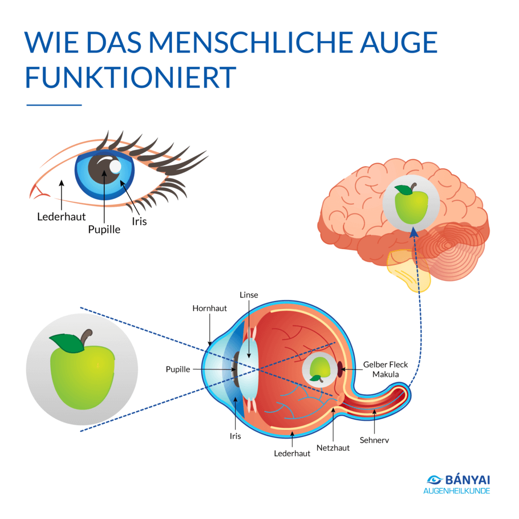
“Seeing” is much more complicated than you might imagine. In order for the environment to become visible, the eye must convert light into nerve stimuli, which are then transmitted to the brain. Only there does the actual picture emerge. The various components of the eye take on very specific and well-coordinated tasks.
In order for you to see an object, the light rays reflected by that object must hit the cornea. There, the light is focused, which then hits the so-called rainbow skin, the iris, behind the anterior chamber of the eye.
Behind the rainbow skin lies the lens. This concentrates the incoming light even more strongly and thus regulates near and far vision. This focused light then passes through the vitreous body behind the lens onto the retina. This creates a preliminary intermediate image, which is upside down and mirrored.
The visual cells convert the light into the actual nerve stimuli, i.e. nerve impulses. Once these impulses have reached the brain via the optic nerve, the finished upright image is created.
Eye whiteness is the visible area of the solid outer shell of the eyeball. The iris resembles a disk with a hole in the middle (pupil) and is the colored area of the eye. It contains muscles that change the size of the pupil. Think of the iris as a camera diaphragm: when it is dark, the round pupil widens. At brightness, this opening becomes smaller. In this way, it controls the amount of incident light to create an optimal image.
The iris and the pupil (the black in the eye) are covered by a translucent layer: the cornea. Your task, together with the eyelids, eyelashes and tear fluid, is to protect the eyes from penetrating foreign bodies or injuries. In addition, the cornea is also involved in the visual process. The incident light rays are bundled here and already refracted.
The cornea does not lie on the iris, but is stretched over it like a small dome. The aqueous humor is located between the cornea and the iris. This is a fluid that cleanses the eye and supplies the cornea and lens with important nutrients.
The light rays that pass through the pupil then strike the lens behind it. This is attached to short muscles via solid fibers, which can change the shape of the lens. The incident light is refracted to different degrees depending on the shape, which allows the eye to adjust to “close” or “wide”. This process is called accommodation.
Behind the lens is the transparent vitreous body (corpus vitreum) inside the eye. Its jelly-like mass gives the eyeball its plump-elastic shape. The fact that the vitreous body is transparent is an important prerequisite for good vision. If there is clouding in old age, eye diseases, such as cataracts, can develop.
The retina is located on the inside of the back wall of the eyeball. In the so-called fundus of the eye – its posterior area – there are millions of sensory cells that receive light stimuli and convert them into nerve signals. The lens refraction creates a sharp, upside-down and upside-down image of the things being viewed.
In the retina, two types of sensory cells are distinguished: cones and rods.
Most cones are located approximately in the middle of the fundus of the eye, the so-called “yellow spot” (macula). This is where the sharpest vision is located.
One of the most common causes of blindness is macular degeneration. In the course of the disease, the retina regresses, resulting in progressive vision loss in the central visual field. A cure for the disease is still not possible. In many cases, however, it is possible to stop or slow down the progression through therapy.
The incoming nerve signals from the cones and rods then travel via the optic nerve into the brain. There, the information is processed and a consciously perceived image is created.1
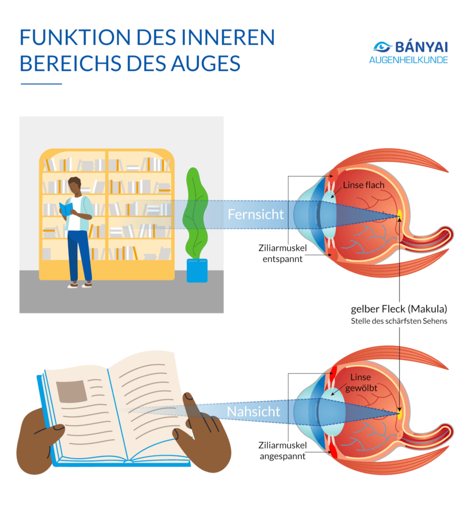
We humans owe our ability to see clearly to accommodation. Accommodation is a muscular process in our eye through which we see sharply in the distance.
The so-called ciliary muscle automatically adjusts the thickness of the lens to the distance of the object. In close objects, the ciliary muscle is relaxed and the lens remains thick. If we look into the distance, the muscle tightens and the lens is pulled apart and flat. This deformation of the lens changes the refractive power of our eyes and objects are automatically focused.
However, the accommodation does not work the same for all living beings. Unlike us humans, for example, dogs tend to be blurry and less coloured. Insects, on the other hand, can even detect ultraviolet light.
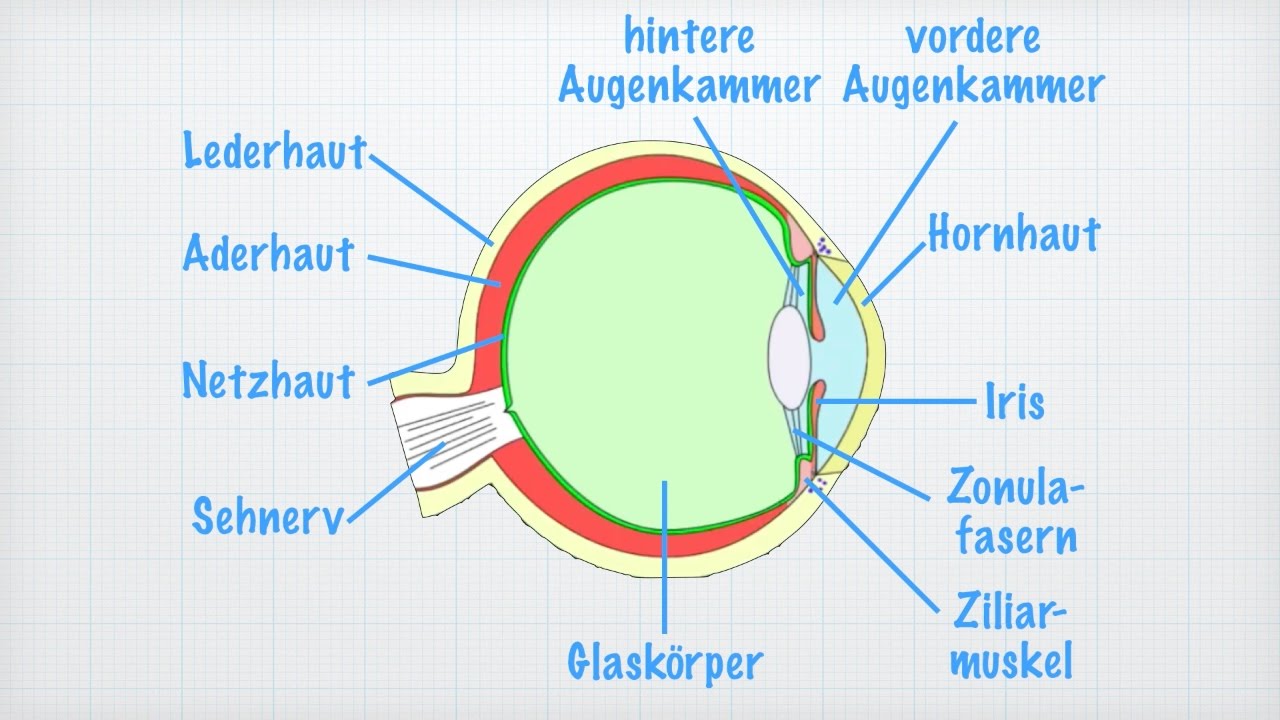
The shape of the eye is a complex anatomical structure. It consists of several structures, all of which perform a certain function. The three most important are:
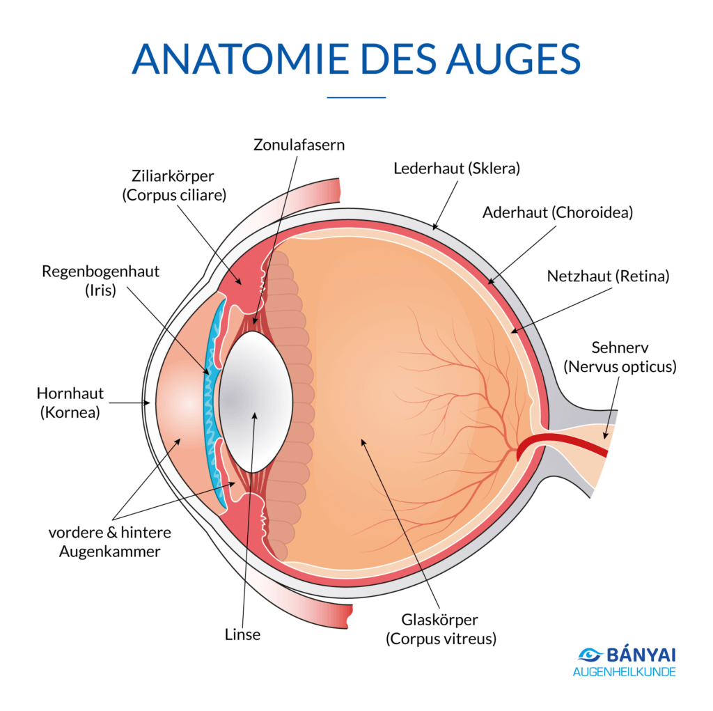
The eyeball is the part of the eye that is also visible from the outside. It probably owes its name to its round shape. In technical language, it is also called Bulbus oculi. The eyeball is covered by three layers – the outer eye skin, the middle eye skin and the inner eye skin. The eyeball is located in the eye socket (orbit).
The outer eye skin is composed of the dermis and the cornea. The dermis gives the eyeball its white color and almost completely encloses it. Only in the foremost area is there a transparent layer – the cornea. Together with the conjunctiva, they serve above all to protect the eyeball from bacteria and foreign bodies.
The middle eye skin consists of the choroid, the ciliary body and the iris. The choroid makes up most of the middle layer and provides the eyeball with nutrients. The ciliary body is responsible for the suspension of the lens. With the help of the pupil, the rainbow skin controls the incidence of light and determines our eye colour.
The inner layer is the retina, which is essentially responsible for vision. There are numerous light-sensing cells in the retina, which are divided into suppositories and rods. These enable us to see colour or recognise black and white tones.
The appendix organs of the eyes include, among other things:
The visual pathway transmits the image from the eye to the brain.
It consists mainly of the optic nerve, which is an approximately 4.5 cm long connection from the eyeball to the brain .
The image is transmitted by more than a million nerve fibres.
ONLINE LASER EYE TEST!
Are you wondering if you’re a good candidate for laser eye surgery? Take our online eligibility test now and find out your results in just a few clicks. Then book your consultation at our laser eye surgery center in Stuttgart or at our laser eye surgery center in Karlsruhe.
About 540 million years ago, the eye began to develop. Eyes as we know them today, however, did not emerge from evolution until 100 million years later. Lens eyes, like those of humans and most vertebrates, are probably the most developed visual organ.
Not all animals have such powerful eyes. Nevertheless, researchers believe that the human lens eye and the less advanced eyes of other animals have the same origin. In addition to the lens or camera eyes, evolution has two other eye types:
The complex eyes and the mirror eyes. The complex eyes of insects or arthropods, which consist of many individual eyes. And the mirror eyes, such as those found in shells, have a reflective mirror that projects the light onto the lens.
Structure of the eye – The human eye is one of the most important and sensitive sensory organs. More than 100 million visual cells convert stimuli into electrical impulses, which run via the optic nerve into the brain and thus enable vision.
External components of the eye such as cornea, dermis, iris and the refractive power of the lens respond to incidence of light. Inside the eye, the stimuli are then processed in the retina. But only your “gray cells” process the incoming nerve impulses and assemble them into an actual image.
Think of the structure and functioning of the human eye as that of a camera. Many individual components work together in perfect harmony in order to obtain a sharp image that is as error-free as possible. In the following, we will explain the structure of the eye and the individual associated anatomical areas.
In the following article, you will learn everything important about the structure and function of the eye and its components.
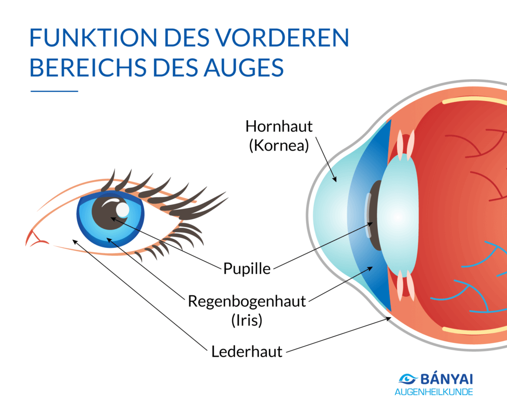
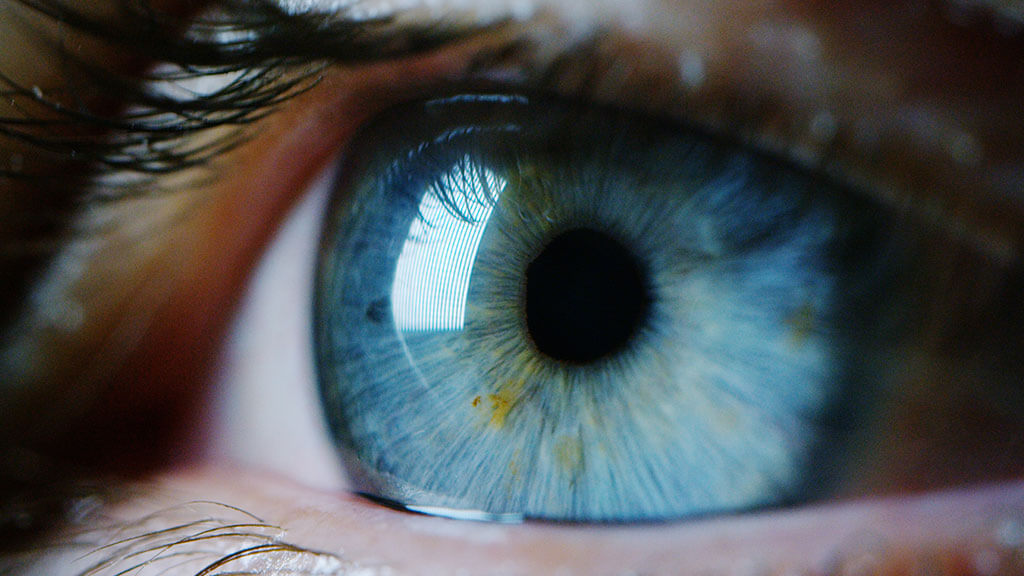
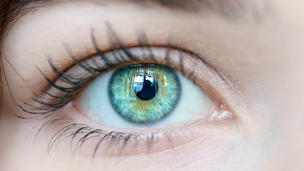
Viewed from the outside, a small black circle first stands out in the center of the eye – the pupil. It is surrounded by the coloured rainbow skin (iris), which gives each person their own individual eye colour. On the outer edge you can also see the white dermis (sclera).
Behind the pupil is the lens. It is not visible from the outside. The lens and cornea – a transparent membrane above the iris – collectively focus the incident light on the retina in the back of the eye. They thus form an important part of the visual process.
Imagine your cornea (lat.: cornea) as a window of the eye: Here the light falls in.
The transparent dome is just over half a millimeter thick and consists of collagen fibers that are finely woven together. Her outside is moistened with tear fluid.
The cornea serves as a protective shield for the eye. It is the transparent, curved front part of the eye and is made up of several layers.
In addition, the cornea, together with the lens, is responsible for refracting the incident light. Incident light rays are focused through the cornea with a refractive power of 43 diopters. This is the only way to create an image on your retina and allow you to see with the eye.
It is traversed by a variety of nerve fibers and covered on the outside by a tear film.
Since the cornea of the human eye is crossed by many nerve fibers, it is very sensitive. Accordingly, a foreign body also feels uncomfortable in the eye. If the cornea is unevenly shaped, this can lead to blurred, distorted vision. This is called a corneal curvature.
The rainbow skin, also called iris, forms the individual, coloured ring of your eye. There are two eye muscles in the iris, which enlarge or reduce the pupil depending on the lighting conditions. The rainbow skin is therefore comparable to the aperture of a camera.
The iris encloses the pupil and separates the anterior and posterior chamber of the eye. It can have different colors as it is colored by pigments. It acts as a diaphragm of the eye, as the muscles of the iris regulate the incidence of light from the eye via the narrowing or widening of the pupil.
Color pigments not only determine your eye color, but also seal the iris against penetrating light. Thus, the light can only enter the eye through the pupil.
The pupil sits behind the cornea, in front of the lens and is enclosed by the rainbow skin. It controls the incidence of light into the interior of the eye by contracting or widening.
The small opening in the middle of your iris is called the pupil. Depending on the prevailing light conditions, the pupil can adjust its radius to create ideal conditions for a sharp image.
In brightness, the pupil is only a tiny opening. In the dark, on the other hand, it widens to allow as much light as possible through. Your state of mind also influences your pupil: if you are excited, frightened or particularly pleased, the pupil can dilate.
A children’s pupil is generally larger than an elderly one.
The white dermis (lat.: Sclera) is the strongest part of your eye. The so-called “sclera” refers to a layer of solid collagen that surrounds the eyeball and protects the eye from injury.
In addition, the dermis helps the eyeball retain its shape. It merges into the cornea at the front. The sclera leaves two gaps at the front for the cornea and at the back for the fibers of the optic nerve.
Since the part of the dermis visible from the outside is covered with transparent conjunctiva, your eye is well protected.
The lens of the eye (lat.: Lens crystallina) is a convergent lens, that is, it focuses the light that enters through the pupil, so that a sharp image is created on the retina. Due to its elastic properties, the lens can adjust its refractive power to focus on both distant and near objects. To do this, it needs the help of a small muscle, the ciliary muscle.
The lens of the eye lies behind the rainbow skin and is surrounded by a fine membrane. It is connected to the ciliary muscle via the so-called zonular fibers. This ensures that the lens can change its shape and thus also its refractive power.
Medically, this sharpness adjustment is called accommodation.
With increasing age, the lens’s ability to accommodate itself decreases, it is no longer as elastic. As a result, the refractive power also decreases and presbyopia occurs.
This form of adaptation makes it possible for you to see both very distant objects and small things directly in front of you. Loss of vision is associated with opacity of the lens of the eye. It is called a cataract.
The shape of the eye resembles a sphere – hence the term eyeball. With a diameter of around 22 millimetres, it is about the size of a 1-euro coin. Three main layers form the basis of the eye: The outer layer consists of the resistant, protective white leather skin. In the anterior region of the eye, it is replaced by the dome-shaped transparent cornea, which takes over most of the refraction of light in the eye.
Under the dermis lies the choroid. It is very well supplied with blood to provide the eyeball with sufficient oxygen and nutrients.
The retina forms the innermost layer. It contains the sensory and nerve cells of the eye and borders inwards on the vitreous body. This fills the inside of the eyeball with gelatinous substance, which is surrounded by a thin shell.
Depending on the position of the layers and components, ophthalmologists refer to the anterior and posterior sections of the eye when setting up the eye.
The chambers of the eye (lat.: Camerae bulbi) have a much smaller extent than the vitreous cavity. They are not filled with a gelatinous mass, but with aqueous humor. This special fluid contains nutrients and oxygen to nourish the lens and cornea on the one hand and stabilize the shape of the eye on the other.
The iris (rainbow skin) delimits the smaller posterior chamber of the eye (lat.: Camera posterior bulbi) forward. In the rear area, the lens connects.
The anterior chamber of the eye (lat.: Camera anterior bulbi) extends from the posterior surface of the cornea to the rainbow skin.
The aqueous humor, which is produced in the posterior chamber of the eye, can be transported through a gap between the lens and the iris into the anterior chamber of the eye. The so-called ventricular angle forms between the iris and the cornea. Here, the aqueous humor can be absorbed through tiny cracks into a small canal, the Schlemm canal, and released into the blood from there.
Depending on age, the intraocular pressure is usually between 10 and 20 mmHg. If the aqueous humor cannot drain properly, the pressure in the eye increases. The result is the so-called glaucoma (green cataract) – a group of different diseases of the eyes in which damage to the optic nerve occurs.
There are two cavities in the front section of your eye, which are filled with colorless aqueous humor. The anterior chamber of the eye usually extends from the cornea to the iris. The posterior chamber of the eye is usually smaller and bounded to the front by the rainbow skin, as well as to the back by the lens.
If the eye chambers are too flat, the aqueous humor cannot drain properly and the intraocular pressure can rise. The result is a cataract, an eye disease that leads to blindness if left untreated.
The vitreous body (lat.: corpus vitreum) fills the inside of the eye between the lens and the retina. The vitreous body occupies the largest part of the eye. It consists of a transparent, gel-like substance and is located between the lens and the retina.
It consists of 98 percent water, in which protein components and the finest connective tissue fibers are distributed, and contains neither nerves nor blood vessels. This gel is surrounded by a boundary membrane, which lies at the front of the lens, and at the back, as well as to the side of the retina.
The high water content is also the reason for its high transparency. The remaining 2 percent is accounted for by hyaluronic acid and collagen fibers. Since hyaluronic acid binds water, the vitreous body has a gel-like consistency.
The eyeball sits behind the lens in the eye socket and is filled by the vitreous. The gel-like substance gives the eye stability and contributes to spherical shape.
The retina contains the sensory cells of the eye (light-sensory cells for color vision and the distinction between light and dark; specialized nerve cells) and is therefore actually an upstream part of the brain. The photoreceptors pick up the light stimulus, process it and transmit the information via the optic nerve to the brain. The retina takes over the function that a film has with the camera.
Although the retina is only about 0.1 to 0.5 millimeters thick, it consists of a total of ten layers. There are a large number of light-sensitive sensory cells in the retina. A distinction is made between two types of light-sensing cells: rods (perception of light and dark; a total of approximately 120 million) and cones (colour vision; a total of approximately 7 million). They are only in the depth of the retina, in the penultimate layer.
In the middle are mainly the cones, which are responsible for colour vision. The chopsticks lie on the edge, which give you a clear view even in the dark.
On each retina there is the so-called macula (macula lutea/yellow spot). It contains a particularly high density of cones and is thus the area of sharpest vision in the eye. The visual pit (fovea), forms the site of the macular center.
The macula is called a “yellow spot” due to its abundantly stored yellow color grains (lutein and zeaxanthin).
In the disease AMD (age-dependent macular degeneration), damage occurs at the site of the sharpest vision.
In addition to a point of sharpest vision, there is also a point on the retina where there are no sensory cells, the so-called “blind spot”. This is the entry point of the optic nerve and the blood vessels that supply the eye.
A rare disease of the eye is retinal detachment. Those affected complain of flashes of light, various visual disturbances. Left untreated, retinal detachment can lead to blindness.
The ciliary body (lat.: corpus ciliare) is an annular bead that surrounds the lens. It contains the also ring-shaped ciliary muscle and therefore contributes to the focusing of objects at different distances. It is connected to the lens via the zonula fibers and adjusts them accordingly (accommodation).
When the ciliary body is relaxed, the zonular fibers are stretched, the lens is flat, and distant things are sharply imaged. If it is tense, the zonula fibers are relaxed, the lens takes on a spherical shape and supports close accommodation (near objects are sharply imaged).
In addition, the ciliary body produces aqueous humor, which fills the space between the lens and the cornea.
A component of the retina is the choroid (lat. Choroidea), which is located on the back wall of the eye. It is separated from the retinal pigment epithelium (RPE) of the retina by the rupture membrane (Rupture Membrane).
The choroidea has the task of supplying the retina with oxygen and nutrients. It forms the middle layer between the dermis and retina and is itself composed of 4 layers.
The thin separation layer allows the transport of nutrients and fluid between the retinal pigment epithelium and the vessels of the choroid. Likewise, metabolic products (“waste”) are transported away via the rupture membrane.
The macula is the area in the center of the retina where most of the visual cells are located. It is located in the middle of the retina and is primarily composed of cones for color vision.
The macula (lat. macula lutea) sits directly next to the junction of the optic nerve, in the center of the retina. Since a lot of the yellow dye lutein is stored in the macula, it is also referred to as the yellow spot. In the middle of this retinal site lies the site of the sharpest vision, the central fovea.
The light receptors (rods and cones) are more densely packed here than anywhere else. If you want to view an object without interference and as sharply as possible, your eyes automatically rotate so that the object is imaged on the fovea centralis of the yellow spot.
If the retina recedes in the area of the yellow spot, this is called macular degeneration. This leads to an increasing loss of vision in the central field of vision and, if left untreated, to blindness.
The blind spot is also called the papilla or mariotte spot. This is where the optic nerve meets the retina. There are no light-sensitive receptors at this point of the optic nerve, which makes the eye blind at this point. In everyday life, people have no restrictions as a result. The blind spot occurs in both eyes, which is why the eye is able to supplement the missing information with the other eye.
The ora serrata refers to the transition from the choroid to the ciliary body, i.e. from the sighted part to the blind part of the retina.
The optic nerve is the first section of the visual pathway that connects the eye to the brain. To finally transmit the information from the retina to the brain, you need the optic nerve (lat.: optic nerve). It is about half a centimetre thick and is formed by a huge bundle of nerve fibres.
The optic nerve exits the retina through the papilla (lat. papilla n.optici), also known as the blind spot. Since there are no light receptors at this point, there is always a small piece missing in the image that the brain perceives.
Under normal conditions, however, you will not notice any of this. Like the retina, the optic nerve is counted as part of the brain.
Diseases of the optic nerve are usually associated with visual field deficits. The optic nerve head is located next to the macula. At this point, the nerve exits the eye.
Upon incidence of light, the light beams are first refracted on the cornea, i.e. deflected. It then passes through the pupil until it reaches the lens. The refractive power of the lens deflects the incident light once again. In this way, it can pass through the vitreous body in the eyeball to the point of sharpest vision in the retina.
The image that arises on the retina is initially upside down.
The light-sensing cells of the retina convert the light into electrical signals that reach the brain via the optic nerve. There it is finally processed and turned back the right way, so that a finished image is created.
The eyelids are mainly responsible for protecting the eyes from external influences. Due to the eyelid closure reflex, we automatically close our eyes before a foreign object or too much light can penetrate the eye.
In addition, the movement of the eyelids distributes the tear fluid, which keeps the eye moist . This prevents the eyes from drying out and small foreign bodies are flushed out via the tear duct.
Often we do not even notice when our eyes lose their vision due to an eye disease. The reason for this is that one eye can compensate for the visual impairment of the other eye. This gives the affected person the impression that they can still see completely. Often, the loss of vision is only noticeable when both eyes are severely impaired. Regular checks at the ophthalmologist therefore help to detect and treat eye diseases at an early stage . You are welcome to book an appointment with Mrs. Doctor-medic Liliana Bányai.
Did you also know that we have two blind spots (also: papillae or mariotte spots) in our field of vision. Where the optic nerve meets the retina, there are no photoreceptors, i.e. no sensory cells that transmit signals to the brain. When you see, you will not notice this, as the blind spots are compensated by the other eye.

Whether short-sightedness or farsightedness, cataracts or greens. Every sufferer of an eye disease is dependent on comprehensive treatment. People have been dealing with the eye and its diseases for thousands of years. But only recently has medicine been able to eliminate refractive errors almost painlessly. In this way, the original visual acuity can be restored almost without exception in a short surgery using an eye laser.
With the right healing method, even serious diseases such as retinal detachment or corneal curvature are detected, treated and vision is improved or maintained.
In most cases, the modern method of laser treatment helps to restore vision completely. Especially in the case of lens opacity, but also in the case of nearsightedness or farsightedness, laser surgery has become a routine procedure. As an alternative to laser in the case of extremely high refractive error or presbyopia, it is also possible to replace the eye lens with an artificial lens.
There are many typical eye problems, the causes of which are usually harmless. If these eye problems persist for several days or the eye pain increases, an ophthalmologist should definitely look at your eyes.

Defective vision refers to a condition of the sense of sight that does not correspond to the norm. Short-sightedness and farsightedness are the most widespread. The eye can no longer see objects either near or far away.
Other forms of refractive error are corneal curvature (also astigmatism or intentionality), night blindness, presbyopia and color vision deficiency.

Affected people cannot see things clearly in the vicinity. This type of refractive error is caused by an eyeball that is too short. Far-sightedness affects fewer young people than older people. Farsightedness that occurs in old age is also called presbyopia.
Things in the distance are blurred in myopia. Here, the eyeball is too long and thus the lens and cornea are too far away from the retina.
As the name suggests, the cornea is deformed in this type of refractive error. The light can therefore no longer be refracted properly. Point-shaped objects are seen by those affected as a line.
People who suffer from night blindness see significantly worse at dusk or in the dark. This is due to a disruption of the light-sensitive cells in the retina.
With this defective vision, some or all shades are not perceived correctly. The reason for this is a dysfunction of the color-sensitive cells in the retina.
One of the most common eye diseases is cataract. This is a clouding of the lens, which usually occurs with increasing age. Green cataract (glaucoma), on the other hand, is an umbrella term for eye diseases that affect the optic nerves.
Other common eye diseases include macular degeneration, keratoconus, retinal detachments or yellow eyes. Regular visits to the ophthalmologist are necessary and recommended in order to diagnose and treat such diseases in good time.
As already mentioned, cataracts are a clouding of the lens. This phenomenon occurs either due to age, inheritance or injury. The impairment can occur either near or far.
The cataract is a disease of the optic nerve in which the optic nerve fibers die. If left untreated, this disease can lead to complete blindness of those affected.
As the name macular degeneration suggests, this disease affects the so-called macula (yellow spot), which is responsible for sharp vision. The progression of vision loss can be stopped by special medications.
A detachment of the retina must always be treated as soon as possible, because in the worst case it leads to blindness. At certain points, the retina detaches from its supply layer. Symptoms of retinal detachment are certain disturbances in the field of vision such as small flashes, shadows or black dots.
Keratoconus is a deformation of the cornea. The cornea bulges conically. This condition is often confused with normal corneal curvature.
The nocturnal cataract is not an actual eye disease, but a clouding of the lens capsule after eye surgery. However, this can be remedied quickly and easily with a laser.
Yellow eyes are usually a symptom of other diseases in the body. They usually occur when the liver is damaged. The most common cause is jaundice.
ONLINE LASER EYE TEST!
Are you wondering if you’re a good candidate for laser eye surgery? Take our online eligibility test now and find out your results in just a few clicks. Then book your consultation at our laser eye surgery center in Stuttgart or at our laser eye surgery center in Karlsruhe.