

ONLINE LASER EYE TEST!
Wondering if you’re a good candidate for laser eye surgery? Take our online eligibility test now and find out your results in just a few clicks.
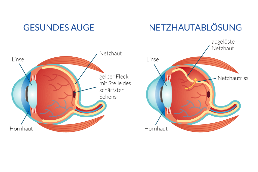
Retinal detachment is a rare and very serious disease of the eye. In ophthalmology, this retinal disease is particularly important because untreated retinal detachment can lead to blindness. The inner part of the retina is detached from its supply layer, the retinal pigment epithelium.
Retinal detachment is an ophthalmological emergency! The first signs of retinal detachment are visual disturbances and the perception of flashes of light. Likewise, visual acuity decreases considerably.
The retina (neuroretina) lies like a kind of skin layer around the vitreous body of the eye and almost completely envelops it. Eye disease such as retinal detachment can lead to painful shrinkage of the eyeball and subsequent blindness. However, this only happens if it is not treated correctly or not at all.
The choroid is vascular and provides the retina with nutrients.
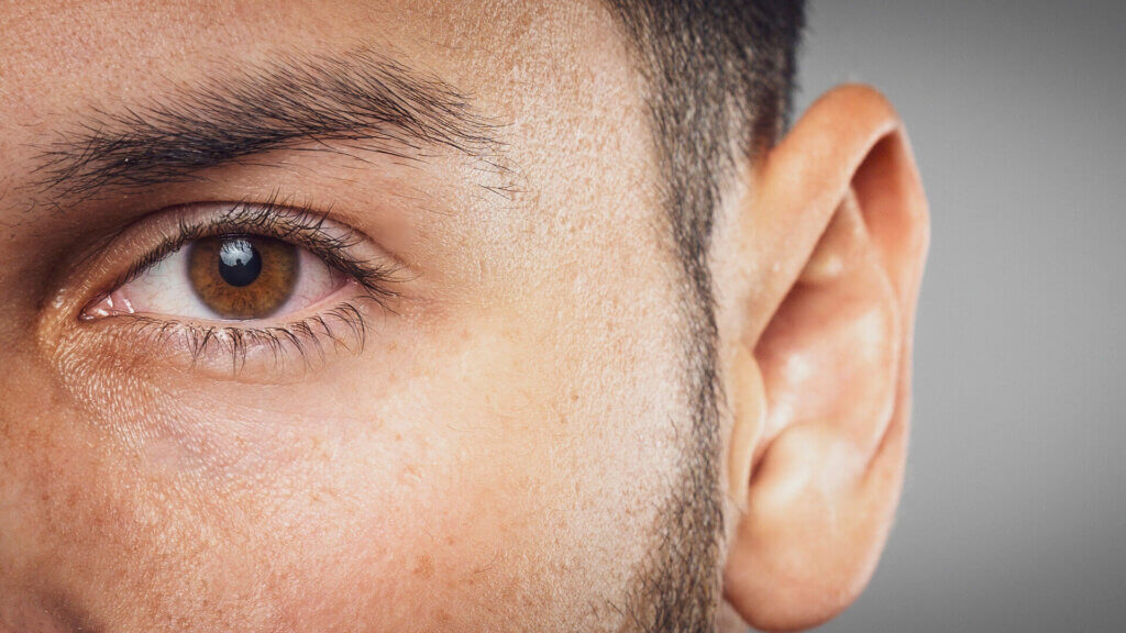
You think they are suffering from retinal detachment? The symptoms of flashes of light in the field of view, black dots, flakes or a black shadow when seeing match? Is your vision deteriorating rapidly? Then you should make an appointment with ophthalmologist Liliana Bányai as soon as possible. Early diagnosis can bring about a quick cure.

Retinal detachment (ablatio retinae and amotio retinae) is caused by the detachment of the two layers from each other. This can have many causes.
In the case of tear-induced retinal detachment, the fluid of the eyeball penetrates between the two layers of the retina via a small tear. This does not necessarily have to lead to a detachment and, as a result, to death. Often, this process can also remain completely symptomless.
In tractive retinal detachment, also called complicated retinal detachment, the upper retinal layer is formally removed by a type of connective tissue in the form of strands inside the eye.
Exudative (fluid-related) retinal detachment is a form of disease in which fluid from the vessels of the choroid passes between the two layers of the retina. This liquid allows the two layers to separate from each other. This form usually occurs due to inflammation of the choroid or tumors.
Very often, a disease of the vitreous body on the retina is the bearer of debt. Inflammation can affect the retina.
Finally, there is the possibility of combining a traction-related rhegmatogenic retinal detachment (retinal tear). In this form, a tear in the retina due to a shrinking vitreous body (vitreous detachment) and the pulling of connective tissue strands inside the eye lead to the detachment of the retina.
ONLINE LASER EYE TEST!
Wondering if you’re a good candidate for laser eye surgery? Take our online eligibility test now and find out your results in just a few clicks.
The retina consists of two superimposed layers that are around 0.1 to 0.5 millimetres thick. One of the layers is called the stratum nervosum and contains the nerve cells. The second layer lies underneath and is called stratum pigmentosum.
There is a wafer-thin liquid-filled gap between the two layers. The negative pressure in this gap holds the two layers of the retina together.
For various reasons, these two layers may nevertheless become detached from each other. This process is called retinal detachment.
As a result of this detachment of the retina, the sensory cells die and cause the aforementioned symptoms, until they go blind. If this detachment also affects the macula (point of sharpest vision), the visual acuity decreases greatly. Read more about the eye!
The following symptoms can be observed with retinal detachment:
The severity of the symptoms always depends on the degree and exact location of the retinal detachment. In any case, it is advisable to consult an ophthalmologist for the symptoms described in order to be able to get to the bottom of the impairment of vision.
If you have such underlying diseases, you should be examined by an ophthalmologist at least once a year in order to be able to quickly detect a detachment, holes or cracks in the retina if necessary.

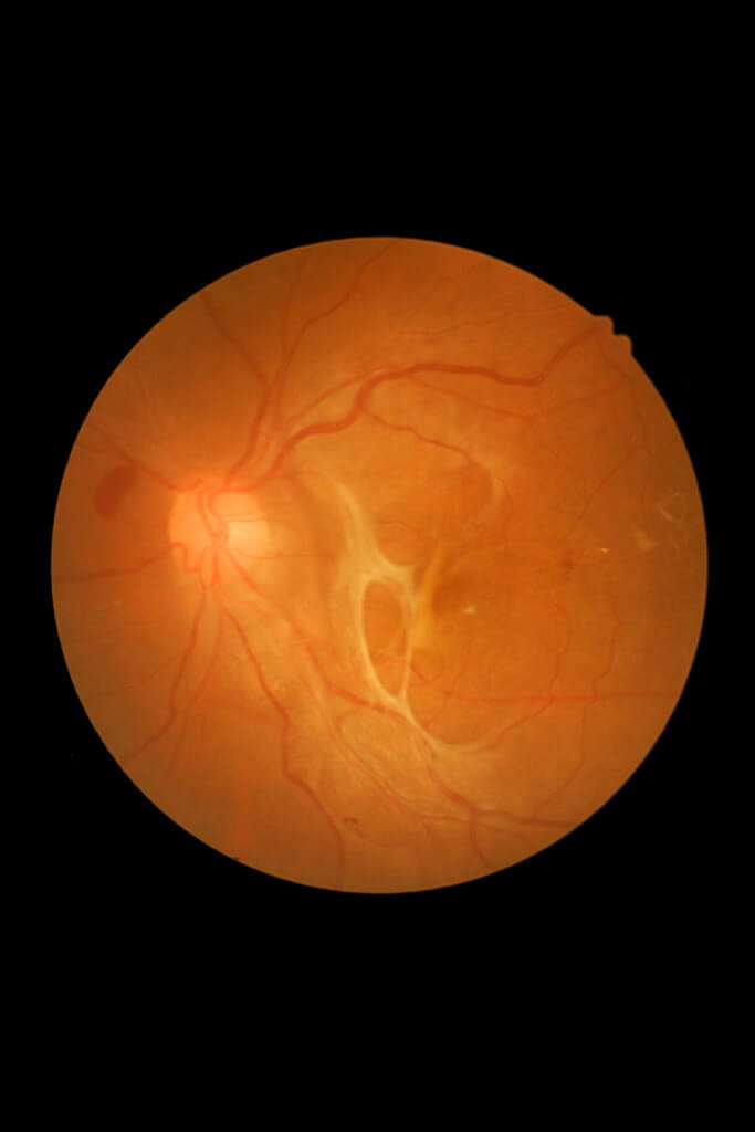
In order to diagnose which form of retinal detachment is involved, a special examination is required. Probably the most important is the so-called eye examination (ophthalmoscopy). By means of a special apparatus (slit lamp), the ophthalmologist is able to examine the fundus. In this way, it recognizes the retina and can immediately recognize any abnormalities such as a hole in the retina.
Bubble-like withdrawals are usually noticeable. The type of damage or its shape can already give an indication of the cause of the detachment of the retina.
In exudative retinal detachment, fluid retention is responsible for the detachment of the retina.
If a clear diagnosis is not possible, the ophthalmologist can also examine the retina using ultrasound.
Retinal detachment is always considered an ophthalmological emergency. If you develop symptoms, you should see an ophthalmologist as soon as possible.
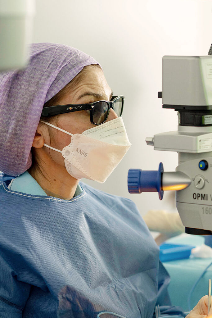
For the treatment of retinal detachment, there is currently no drug option and surgery in an eye clinic is necessary. The eye is usually treated with a laser. Laser treatment can correct the detachment of the retina.
Preventive measures for retinal detachment are annual checks with the ophthalmologist. There, a hole or holes in the retina can be diagnosed early in the healthy eye. Rapid treatment of these retinal holes is advisable.
There are several forms of retinal detachment treatment. One option is laser treatment. The two retinal layers are reconnected by means of a special laser (photocoagulation) or a cold probe (cryopexy). Where the laser or the cold probe strikes the retina, a small scar is created, which connects the skin layers again, but will still impair vision a little.
Another method is the insertion of the eyeball from the outside by means of eye surgery. In this case, pressure is applied to the outermost layer of the eyeball (dermis) by a seal or cerclage during surgery and local anaesthesia, which presses the upper layer back against the lower layer. This operation takes between 30 and 60 minutes. This procedure can take place under local anaesthesia or under general anaesthesia.
The most recent way to counteract retinal detachment is to remove the vitreous (vitrectomy). This operation can also be performed under local anesthesia and takes between 30 and 60 minutes on average.
In this method, the liquid of the vitreous body is sucked off and replaced by a special liquid (silicone oil, gas or Ringer’s solution). This displaces the accumulated fluid between the skin layers and thus joins them together again.
Book your personal consultation online now—with no obligation!
Laser Eye Surgery, Diagnostics & Consultations on Ophthalmology at the Bányai Laser Eye Center – Dr. Liliana Bányai

