
ONLINE Glaucoma Surgery Eligibility Test
Are you wondering if you’re a good candidate for glaucoma surgery? Take our online aptitude test now and find out your results in just a few clicks.
Glaucoma diagnostics is based on more than just measuring intraocular pressure (IOP). In order to rule out irreversible damage to the optic nerve, modern imaging methods are indispensable.
Optical coherence tomography (OCT) and visual field measurement (perimetry) provide high-resolution data and functional evidence. Only the combination of these procedures provides you and our team of experts with the necessary security for early treatment.
Modern Green Star diagnostics goes far beyond the mere measurement of intraocular pressure (IOP). It is indispensable because IODINE alone does not have sufficient information about the health of your optic nerve.
Only through the combination of imaging methods (OCT) and functional tests (visual field measurement) can we detect damage at an early stage and thus prevent irreversible vision loss. We value this comprehensive, technologically advanced investigation.
The measurement of intraocular pressure (IOP) is only a first indication. Many patients with normal pressure glaucoma have IOP values that are within the normal statistical range. Nevertheless, her optic nerve is damaged unnoticed.
A treatment that is exclusively fixed on the pressure value therefore carries a high risk. Only the imaging analysis using OCT shows us the actual state of the optic nerve and the retina.
We recommend the comprehensive “Eye Check 40 Plus” every year from the age of 40. This package includes the macular and glaucoma examination using OCT.
In addition, we perform the visual field measurement, the applanation tonometry and the representation of the entire posterior segment of the eye. The aim is to detect eye diseases such as incipient damage to the optic nerve at an early stage and thus avoid irreversible vision loss.

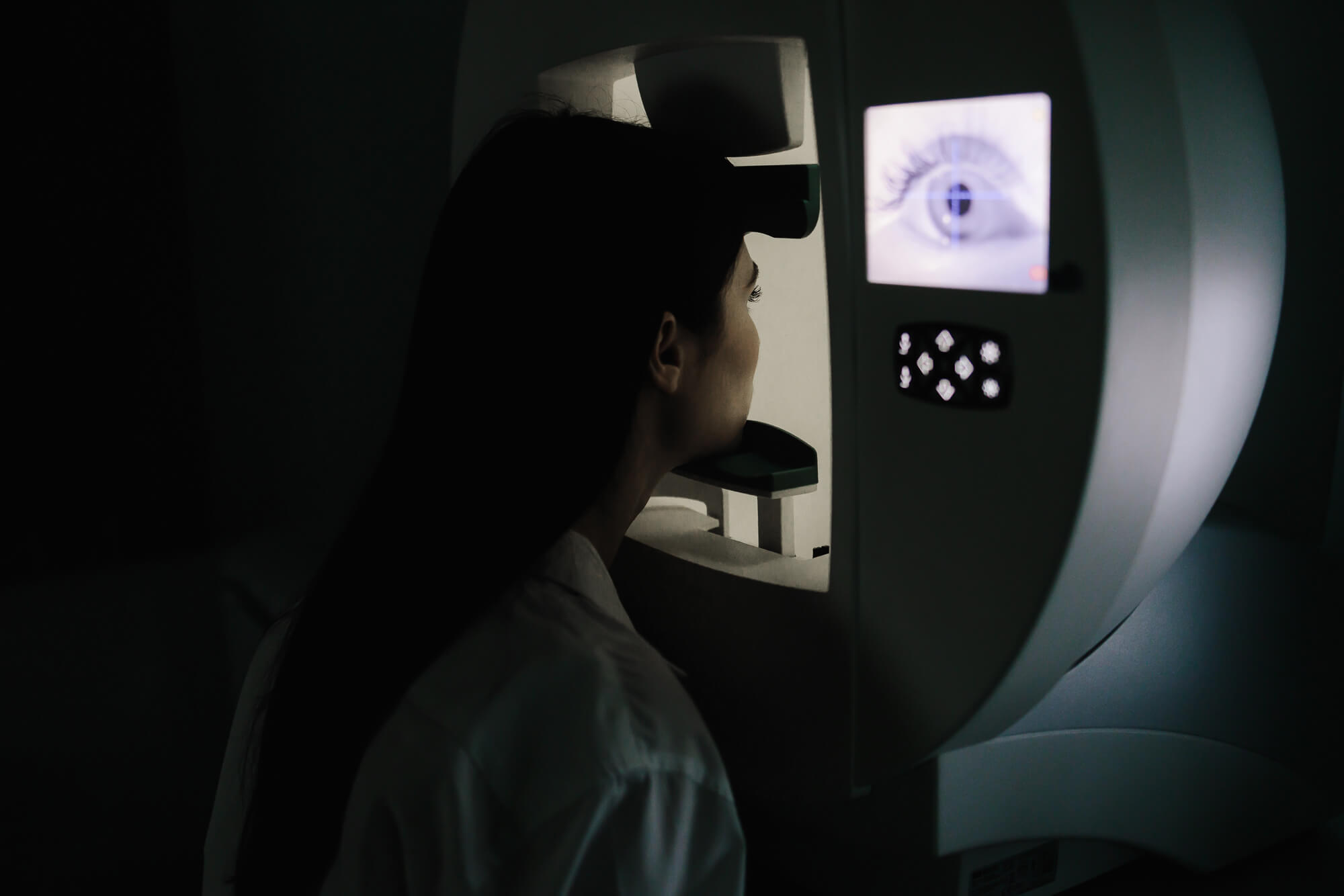
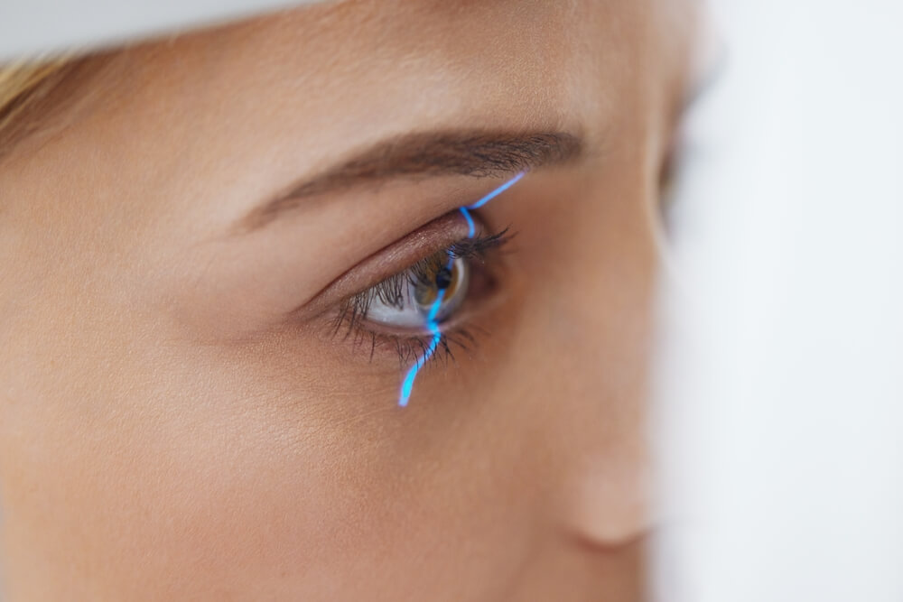
Optical coherence tomography (OCT) is a key technology in Glaucoma diagnostics. It enables a high-resolution representation and documentation of the internal structures of the eye.
The procedure is fast, non-invasive and provides important information about the thickness of the retinal nerve fiber layer (RNFL) and the structure of the optic nerve head. This detailed view is crucial for detecting even the smallest, glaucoma-typical damage at an early stage.
Glaucoma OCT is an imaging examination that captures the structure of the optic nerve head and retina.
With high-resolution images, it makes it possible to detect the smallest changes caused by the cataract at an early stage. This is essential because the damage to the optic nerve is irreversible. An early diagnosis by the OCT allows us to initiate immediate therapy.
In addition to glaucoma-specific OCT, macular OCT is also of great importance. It is a specialized form of Oct examination that details the area of the macula (the sharpest point of vision).
This enables early detection of changes, such as age-related macular degeneration, and precise treatment planning. Both Oct forms ensure comprehensive diagnostics.
ONLINE Glaucoma Surgery Eligibility Test
Are you wondering if you’re a good candidate for glaucoma surgery? Take our online aptitude test now and find out your results in just a few clicks.
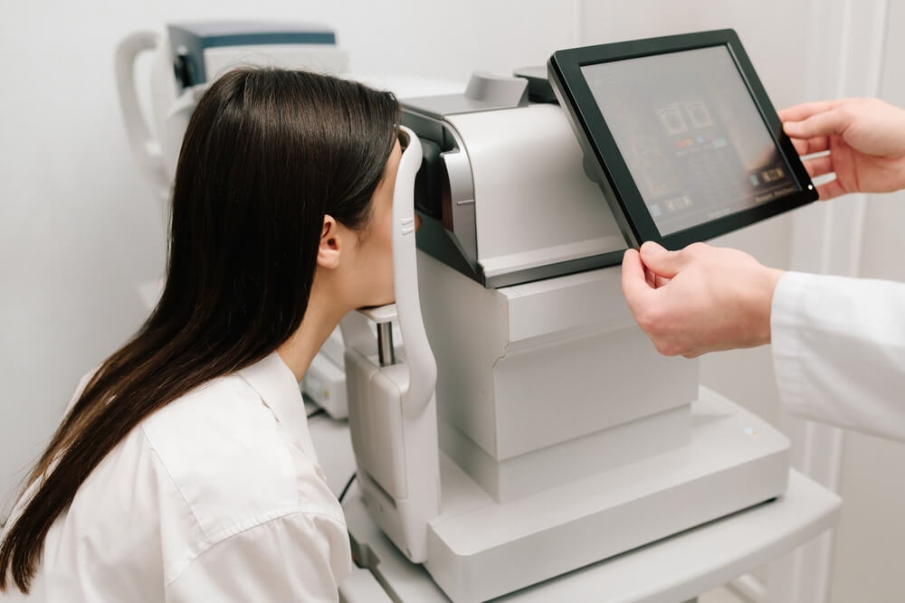
Visual field measurement (perimetry) is the functional test in glaucoma diagnostics. It checks how well patients perceive objects in different areas of their field of vision.
This measurement is crucial to determine possible malfunctions or limitations. It provides direct evidence of how far the disease has already affected peripheral vision.
Perimetry projects points of light of varying intensity as you look straight ahead.
The test only takes a few minutes and is painless. The measurement is carried out to detect diseases such as glaucoma or retinal diseases. The results show us which parts of the visual field have already failed – this correlates with the damage to the optic nerve that becomes visible in the OCT.
We attach great importance to comfortable and efficient glaucoma diagnosis and examination. Unlike older procedures, we do not require pupil dilation, which saves you time and maintains your ability to drive.

The following procedure and several methods are often only fully reimbursed by private insurance companies. Statutory health insurance companies often do not cover individual benefits. However, the examinations are crucial to make a precise diagnosis and correctly determine the target pressure. We will be happy to provide a cost estimate on request.
1. Preliminary examination:
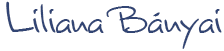
Refraction (spectacle lens power), visual performance and intraocular pressure (applanation tonometry).
2. Field of view measurement (perimetry):
Functional test of the field of vision; detects early limitations due to glaucoma or retinal diseases.
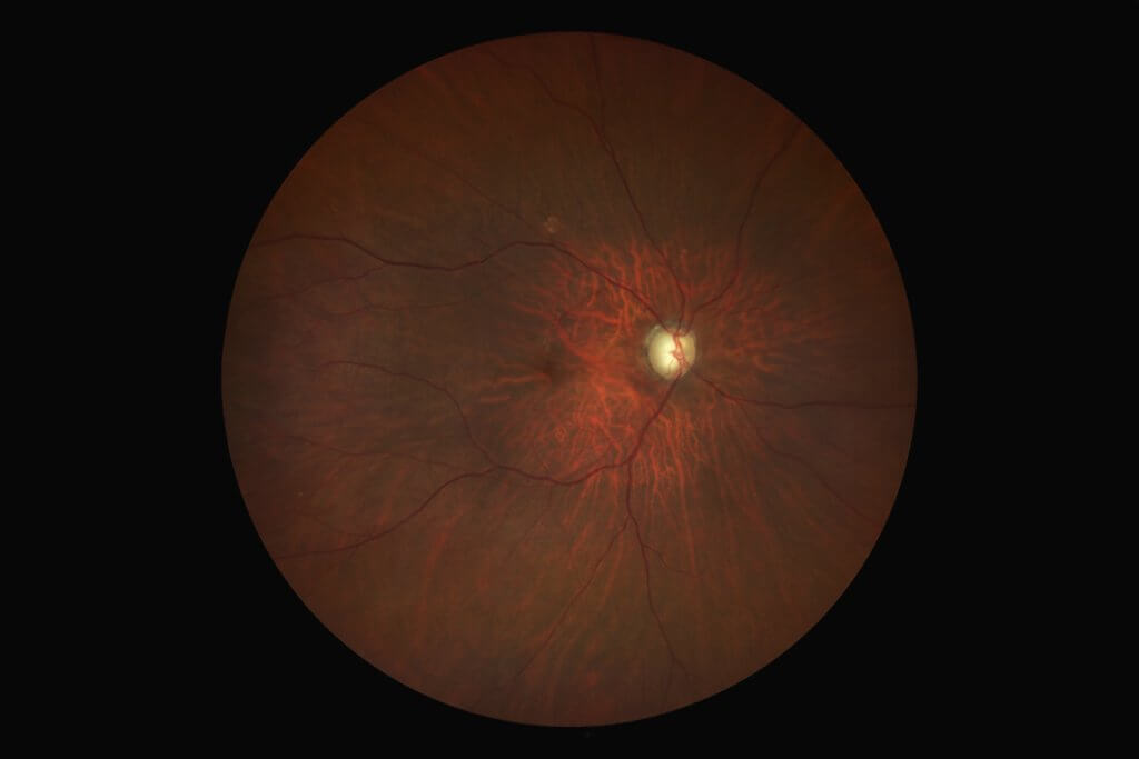
3. Eye fundus without pupil dilation:
High-resolution fundus images from the macula to the periphery with a modern retinal camera – true to colour, gradual, mobile.
4. Optic nerve examination / glaucoma OCT:
Imaging of optic nerve head and RNFL in high resolution; shows glaucoma-typical changes early.
5. Macular OCT:
Detailed representation of the macula (e.g. in age-related macular degeneration); supports therapy planning.
Recommendation: Carry out annually as a precautionary measure from the age of 40 (“Eye Check 40 Plus”). Services and reimbursement depend on your private tariff.
We use technologically advanced retinal cameras to view and document the entire posterior segment of the eye. Without pupil dilation, high-resolution fundus images are recorded from the macula to the periphery of the retina.
These color-accurate images simplify the diagnosis and documentation of eye diseases and offer unprecedented certainty in the diagnosis.
Book your personal consultation online now—with no obligation!
Laser Eye Surgery, Diagnostics & Consultations on Ophthalmology at the Bányai Laser Eye Center – Dr. Liliana Bányai

Comprehensive glaucoma diagnostics with OCT and visual field is one of the individual health services (IGeL). The statutory funds often do not take over these preventive examinations, as they are not part of the standard benefits.
At Bányai Neue Augen, however, we are convinced that these preventive measures are essential for the preservation of your eyesight.
The cost of OCT and visual field is an investment in the long-term preservation of your vision. They enable the early detection of damage before it affects central vision.
In this way, a Green Star operation or a massive loss of vision can be avoided. Advanced diagnostics support treatment by the ophthalmologist.

Detecting earlier means treating better. With the “Eye Check 40 Plus”, you get a precise diagnosis before damage becomes noticeable – and thus the decisive advantage for your eye health.
The comprehensive screening, including intraocular pressure measurement, visual field and OCT, is designed to be efficient and time-saving. Because we use modern retinal cameras, pupil dilation is not required. The entire process for creating an accurate diagnosis usually takes only about 30 to 45 minutes.
No, the detailed examinations using OCT and the visual field measurement are extended preventive services. They are offered to legally insured persons as Individual Health Services (IGeL). As a rule, statutory health insurance companies do not cover the costs of these modern diagnostic procedures.
Corneal thickness is an important correction factor in intraocular pressure measurement. Measuring the IODINE is only meaningful in conjunction with measuring the thickness of the cornea. A cornea that is too thick or too thin can falsify the measured pressure. Measuring the thickness of the cornea is therefore essential for accurate glaucoma diagnosis.