
ONLINE LASER EYE TEST!
Wondering if you’re a good candidate for laser eye surgery? Take our online eligibility test now and find out your results in just a few clicks.
This term from ophthalmology comes from the Greek composition: keratos for “horn” and cone for “conical”.
Keratoconus is a pathological process that affects the eye. During keratoconus, a progressive thinning and deformation of the cornea occurs. This results in a change in the thickness of the cornea. The cornea can deform so much that it takes on a conical shape. The inner corneal layer may also tear. Keratoconus is untreated, i.e. a progressive corneal disease.
Keratoconus occurs between the ages of 20 and 30. The cornea can develop and deform in keratoconus patients up to the age of 40 or 50. In the initial stage of conical protrusion, soft contact lenses or spectacle lenses can still help to compensate for the irregularities of the cornea and maintain visual performance.
It happens again and again that the disease keratoconus is not recognized and is confused with a common curvature of the cornea of the eye (astigmatism).
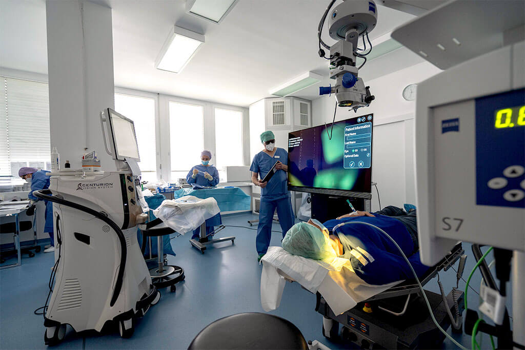
Thanks to our advanced technology and years of experience, we can determine exactly whether or not you suffer from a disease of the corneal tissue – keratoconus. In the course of examining and diagnosing keratoconus, we can initiate ways to treat keratoconus.


The disease keratoconus appears in two different forms:
The silent form of keratoconus is usually indistinguishable from the normal curvature of the cornea and is usually only detected by a close examination of the eyes. This form of keratoconus is the most common and occurs ten times more often than the progressive form. Depending on the stage, contact lenses and glasses help.
If the patient suffers from progressive keratoconus, cracks and thinning of the cornea may occur. This can damage the cornea to such an extent that only a corneal transplant (perforating keratoplasty) can help.
ONLINE LASER EYE TEST!
Wondering if you’re a good candidate for laser eye surgery? Take our online eligibility test now and find out your results in just a few clicks.
The causes of keratoconus can be quite different and have not yet been fully clarified. It is suspected that there is a disruption between the collagen molecules of the connective tissue and the cornea. There is also the possibility of genetic transmission of keratoconus, as this eye disease of the cornea often occurs in several family members.
Increasingly, this disease of the cornea also occurs in patients with trisomy 21. This is better known as Down syndrome and affects between 30,000 and 50,000 people in Germany.
Another assumption is that this disease of the cornea is increased in allergy sufferers, as they rub the eye more and this probably promotes the development and progression of keratoconus.

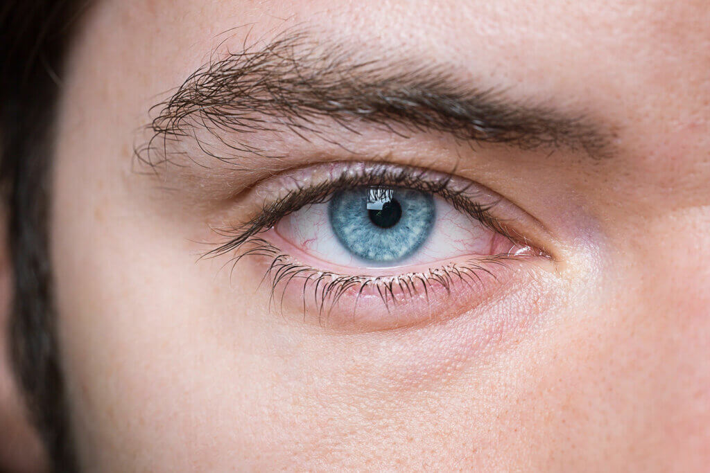
One of the most common symptoms of keratoconus is the fluctuation and decrease in visual acuity through the cornea – a deterioration in vision. Increased aperture and light sensitivity is also often noticeable. This often leads to so-called halos and you perceive light rings around a light source.
If the corneal change progresses further and further, short-sightedness usually develops. Measuring the surface of the cornea is an important part of diagnostics.
If you experience such symptoms of keratoconus, it is advisable to visit the ophthalmologist and undergo a more detailed examination. Read more about the eye!
In the treatment of keratoconus, the shape of the corneal disease plays a crucial role.
In the silent form of keratoconus, glasses or contact lenses are usually sufficient for correction. Likewise, dimensionally stable contact lenses can be used to treat the cornea.
In the progressive form of the disease keratoconus, contact lenses can only help in the initial form. As the disease progresses, the shape of the cornea changes and a contact lens or glasses become obsolete. An effective method of treating the cornea at our eye surgery centre in Stuttgart is so-called UV riboflavin crosslinking, or CXL for short.
Crosslinking is another method of treating keratoconus. When cross-linking, the cornea of the affected eye is saturated with special eye drops (vitamin B2) and then treated with UV radiation. The fibres of the cornea are roughened and the cross-linkages of the cornea are reinforced. The aim of cross-linking is to prevent the progression of keratoconus through these strengthened cross-links. After cross-linking, the keratoconus can no longer progress, but the patient still needs glasses.
Laser eye surgery for keratoconus is therefore the better option if you still want to do without vision aids such as glasses or contact lenses.
Book your personal consultation online now—with no obligation!
Laser Eye Surgery, Diagnostics & Consultations on Ophthalmology at the Bányai Laser Eye Center – Dr. Liliana Bányai

The word keratoconus is a compound word from Greek and Latin. The word Keras (Greek) means horn. The Latin word conus means cone. Keratoconus means shortly, corneal cone.
This refers to a progressive thinning – as well as a protrusion of the cornea.
In the disease of keratoconus, you basically see the same as with a corneal curvature. One suffers from stubbornness, as well as short-sightedness or farsightedness. The special feature of keratoconus, however, is the constant deterioration of the field of vision through the cornea, in which vision rapidly decreases despite frequent changes of glasses.
The treatment of the disease keratoconus, a patient is usually paid by the health insurance, but in certain cases not: If patients need financing, this is also possible with us. To learn more about the treatment of keratoconus, it is advisable to arrange a consultation with us.
Patients cannot go blind due to keratoconus. In most cases, patients can continue to lead a normal life with the help of glasses or hard contact lenses. Only about 10% to 20% of patients require corneal surgery.
Acute keratoconus is an ophthalmological (= affecting the eye) emergency. This is a strong clouding of the lens. Patients must be treated immediately at this stage. Glasses or contact lenses no longer help.
