
The early detection and monitoring of glaucoma are based on a combination of several examination methods. Two methods are particularly important: optical coherence tomography (OCT) and visual field testing (perimetry).
But how do these procedures differ? And when is each examination appropriate?
In this post, Doctor-medic Liliana-Iulia Bányai of Bányai Augenheilkunde explains the differences in a clear, practical, and well-founded manner.

Glaucoma is a chronic eye disease in which the optic nerve is gradually and usually painlessly damaged.
The most common risk factor is elevated intraocular pressure, but damage can also occur even at normal pressure.
The insidious part: Many people affected don’t notice the condition for a long time, as the loss of vision develops gradually—usually beginning at the edge of the visual field.
If glaucoma is not detected and treated early, it can lead to progressive vision loss, even blindness. That is why regular preventive check-ups are crucial.
Two of the most important diagnostic procedures are optical coherence tomography (OCT) and visual field testing (perimetry). But what distinguishes them, and when is each test appropriate?
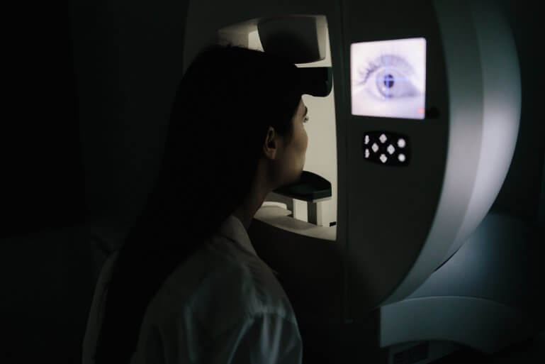
OCT (optical coherence tomography) is a high-resolution imaging technique that uses light waves to produce microscopically precise cross-sectional images of the retina, particularly of the optic nerve head and the retinal nerve fiber layer (RNFL).
This enables a precise assessment of the structures that are first damaged in glaucoma.
Unlike many other examination methods, OCT is completely non-contact, quick, and comfortable for patients.
OCT is particularly valuable because it can often reveal structural changes even before symptoms or functional loss occur. This makes it an early warning system in glaucoma diagnostics.
In addition, OCT enables precise documentation of changes over time.
In this way, it can be assessed whether an existing therapy is having the desired effect or whether adjustments are necessary. In follow-up monitoring, it therefore provides valuable objective data.
Perimetry is one of the functional tests in ophthalmology. It measures vision across the breadth and depth of the visual field and assesses how well the optic nerve transmits visual information from different areas of that field.
Patients look at a central fixation point while light pulses must be perceived from different angles.
This examination is essential for detecting functional impairments that patients often notice only at a late stage — for example, small peripheral visual field defects.
With glaucoma, symptoms often appear gradually, so the actual loss of vision is frequently underestimated.
Visual field testing requires concentration and active participation, so it is of limited use in some patient groups – such as children or very elderly patients.
Nevertheless, it remains an indispensable tool for assessing vision.

The OCT reveals changes in the structure of the optic nerve and retina—particularly thinning of the nerve fiber layer—while the visual field examination maps the impact of these structural changes on vision.
Glaucoma can show only structural changes at an early stage—these are detected by OCT. Only later, once optic nerve tissue has been lost, do visual field defects appear.
Conversely, functional limitations can also occur even if the OCT does not show significant abnormalities — for example, fluctuating pressure readings or other risk factors.
Therefore, in modern glaucoma screening, only the combination of OCT and perimetry provides a complete picture. The two methods are not competitors but complement each other optimally—both for diagnosis and for monitoring disease progression.
The following overview indicates which examinations are particularly useful, depending on the findings and the stage of glaucoma:
| Glaucoma status | OCT (structural diagnostics) | Visual field (functional diagnostic testing) |
| Suspected glaucoma | Highly recommended (early warning sign of damage) | Optional, if early symptoms are present. |
| Initial diagnosis | Standard examination to assess the nerve fibre layer | Indispensable for assessing functional impairment. |
| Follow-up | regular monitoring of structural changes | regularly to monitor changes in vision |
| Change of treatment | to evaluate the efficacy on the optic nerve | to determine whether the visual field has changed |
| limited cooperation | particularly suitable (objective measurement) | Possible to a limited extent (requires concentration) |
| Subjective visual symptoms despite an unremarkable OCT | Additionally useful for identifying functional deficits | especially important |
The combination of OCT and visual field testing is essential in glaucoma screening and treatment.
It enables us to identify risks at an early stage, tailor therapies precisely, and provide individualized care to patients.
At Bányai Augenheilkunde, we use the latest technology and extensive experience to preserve your vision over the long term.
We routinely use both methods in glaucoma diagnostics, during follow-up examinations, and for treatment monitoring, tailored to your personal risk profile.
Early detection means better treatment. And targeted treatment means preserving vision.
No, OCT is a completely non-contact and painless procedure. It uses light waves, not radiation, and can therefore be repeated as often as necessary — ideal for follow-up examinations.
Perimetry takes about 5 to 10 minutes per eye, depending on the type of test. It is important that you remain focused during the examination and keep your eyes fixed on the fixation point.
For stable glaucoma, we generally recommend an OCT and visual field test once or twice a year. If there are active findings or changes in therapy, more frequent check-ups may be necessary – we determine this on an individual basis.
A single test is not enough. Only the combination of several examinations (including measurement of intraocular pressure, OCT, visual field testing, and optic nerve assessment) allows a reliable diagnosis or the exclusion of glaucoma.
We discuss the findings with you in detail, set an individual target for intraocular pressure, and jointly develop a treatment plan. Depending on the findings, more frequent monitoring, therapy adjustments, or further diagnostic testing follow.
Hat Ihnen dieser Beitrag gefallen? Teilen Sie ihn mit anderen!