
Intraocular pressure — medically abbreviated IOP and also called tensio — is a central measurement in ophthalmology.
But what exactly does this value indicate? When is it considered elevated? And what does that mean for the risk of glaucoma (green star)? We at Bányai Augenheilkunde clearly explain what matters in the assessment and why it’s worth knowing your eye measurements.
The normal intraocular pressure in most people is between 10 and 21 mmHg (millimetres of mercury). It results from the aqueous humour in the eye – a clear fluid that is continuously produced and drained through fine channels.
If this balance is disrupted, pressure can build up inside the eye. This pressure regulation is essential for nourishing the internal structures of the eye and for maintaining vision.
If the measured pressure exceeds 21 mmHg, specialists refer to it as an elevated intraocular pressure. If this persists without any detectable damage to the optic nerve or visual field, it is referred to as ocular hypertension.
However, this does not necessarily mean that a disease such as glaucoma is present. What matters is how sensitive the optic nerve is to pressure.
Some people tolerate slightly elevated eye pressure well, while others can develop damage even at “normal” pressure (normal-tension glaucoma). Therefore, an individual assessment by an ophthalmologist is indispensable.

A persistently elevated intraocular pressure is one of the most important risk factors for glaucoma — a chronic condition in which the optic nerve is progressively damaged.
If left untreated, this can lead to visual field loss and, in the worst case, blindness. The good news is that if glaucoma is detected early, its progression can often be halted.
Early diagnosis and targeted treatment—medication-based, laser-based, or surgical—can protect the optic nerve and preserve quality of life.
A glaucoma diagnosis is not based on a single measurement but on a combination of several tests.
Tonometry measures intraocular pressure, either non-contact using an air puff or more precisely with applanation tonometry. This method is performed under local anesthesia and provides particularly reliable measurements.
In addition, OCT (optical coherence tomography) is used. It enables high-resolution imaging of the nerve fiber layer and the optic nerve head.
This makes it possible to detect even the smallest structural changes early – before any functional limitations become noticeable.
Another component is the visual field test (perimetry). This test checks whether and where the visual field is restricted.
Many patients do not initially notice such deficits, and perimetry provides objective evidence of early or progressive damage.
In addition, individual factors such as corneal thickness (pachymetry), diurnal pressure fluctuations, and the course observed across multiple follow-up appointments are taken into account in the assessment. Only by combining these elements can a reliable picture of eye health be formed.
The goal of any glaucoma treatment is to lower intraocular pressure permanently and relieve pressure on the optic nerve.
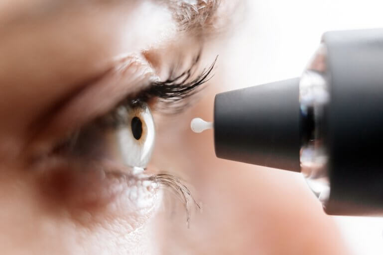
Therapy often begins with eye drops. These either reduce aqueous humor production or improve its drainage.
Depending on the individual case, different classes of medication may be used – such as prostaglandin analogues, beta-blockers, or carbonic anhydrase inhibitors.
Laser treatment can be beneficial for certain types of glaucoma. Selective laser trabeculoplasty (SLT) is an established procedure that specifically targets the trabecular meshwork in the eye and thereby improves the outflow of aqueous humor. SLT can be used as an adjunct to, or as an alternative to, eye-drop therapy.
If this is not sufficient, surgical procedures offer an effective solution. The classic trabeculectomy creates an artificial drainage pathway for the aqueous humor.
In many cases, minimally invasive glaucoma surgery (MIGS) or combined cataract surgery is also considered — especially if lens opacity is present at the same time.
These procedures not only lower pressure but also often improve the quality of vision.
In addition to medical treatment, lifestyle factors should not be overlooked.
Reducing stress, getting enough sleep, exercising regularly, and taking prescribed medications as directed support successful treatment.
We recommend regular eye examinations—especially starting at age 40 or if you have known risk factors such as:
The earlier abnormalities are detected, the more effectively risks can be minimised. The measurement is quick and painless and provides important information for prevention.
To better interpret the measurements, here is an overview of typical pressure ranges and their significance:
| Intraocular pressure | Meaning |
| less than 10 mmHg | Rather low; may require further evaluation. |
| 10–21 mmHg | Normal range |
| greater than 21 mmHg | Indication of elevated pressure, possibly ocular hypertension. |
| 25 mmHg | Significantly elevated; glaucoma suspected. |
Increased intraocular pressure often causes no noticeable symptoms — this is what makes glaucoma so insidious.
The initial symptoms of glaucoma—blurred vision, halos around lights, or visual field defects—often don’t appear until permanent damage to the optic nerve has already occurred.
Important to know: measuring intraocular pressure in isolation is only one part of glaucoma diagnosis.
Equally crucial are the assessment of the optic nerve, the visual field measurement, and imaging methods such as OCT (optical coherence tomography).
In our practice, we combine modern technology with individualized risk analysis – to ensure the highest standard of eye health. Corneal thickness (pachymetry) also plays a role, as it can affect measurement results.
Whether for glaucoma prevention or as part of general eye health — those who know their eye measurements can take action early.
Have your intraocular pressure checked regularly, and discuss any abnormal readings with your ophthalmologist.
One thing is clear: when it comes to glaucoma, every early detection matters. Acting early can help preserve eyesight permanently — for a life with clear vision.
Not necessarily. Elevated intraocular pressure (ocular hypertension) can be a risk factor for glaucoma, but it does not automatically indicate damage to the optic nerve. The key factor is an individual’s pressure tolerance. That is why regular monitoring and comprehensive diagnostic testing are so important.
We recommend an ophthalmological examination every one to two years from the age of 40 — earlier and more frequently if known risk factors are present, such as a family history, diabetes, high myopia, or high blood pressure. Only through regular examinations can changes be detected early.
Yes. In addition to regular eye check-ups, a healthy lifestyle, adequate sleep, stress-reduction measures, a balanced diet, and regular exercise all support eye health. Consistent use of prescribed eye drops is also essential for glaucoma management.
Hat Ihnen dieser Beitrag gefallen? Teilen Sie ihn mit anderen!